Hospitals and health systems
One imaging record across departments, campuses and clinical teams.
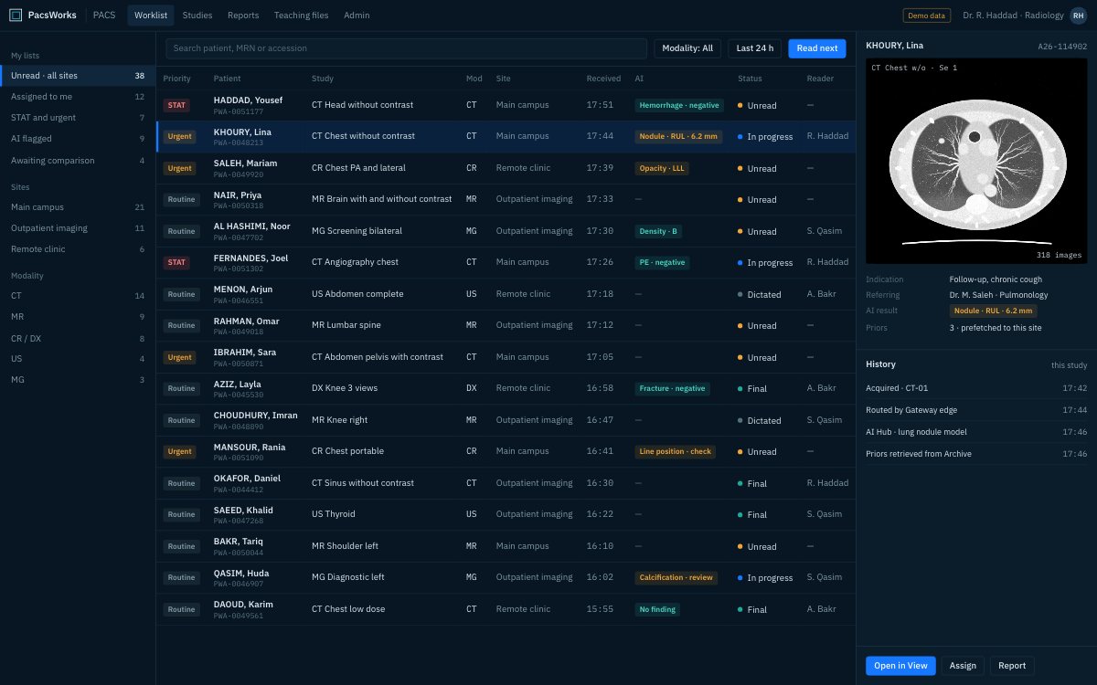
One worklist for every campus, with priority, modality, site and AI flags side by side. Screens show demo data and synthetic images.
The challenge
Hospitals run imaging across radiology, cardiology, emergency and outpatient services, often on several systems at once. PacsWorks brings them onto one platform without a single disruptive cut-over.
Few hospitals chose their imaging estate in one go. Radiology bought a PACS, cardiology bought another, a new campus arrived with its own, and an acquired clinic brought a fourth. Each holds part of the patient’s imaging history, and each has its own contract, upgrade cycle and administrator.
The cost shows up in the reading room and on the wards. Priors sit in a different system, emergency studies queue behind routine work, and clinicians ask radiology to burn discs or send screenshots because they cannot reach the images themselves.
PacsWorks consolidates that estate step by step. A vendor-neutral archive becomes the long-term home for every department’s images, one worklist spans every campus, and clinicians open images from the EMR in a browser, while each legacy system keeps running until its data has been moved and reconciled.
How it fits together.
Every scanner sends through a gateway into one PACS and one archive. Reading, clinical access and the recovery copy all draw on the same record.
An emergency head CT, end to end.
- Scanner
Acquired
The emergency CT sends the study to the edge gateway as soon as the scan ends.
- Gateway
Routed
Rules send it to the PACS, the archive and any head-CT model connected to AI Hub.
- AI Hub
Prioritised
A positive AI result moves the study to the top of the emergency worklist, with a flag.
- Radiologist
Read
The on-call radiologist opens it with priors already loaded and confirms or rejects the finding.
- EMR
Shared
The signed report returns over HL7 and the emergency physician opens images and report from the patient record.
What changes.
Consolidate PACS across sites
Bring separate departmental or site PACS onto one platform, one site at a time.
An enterprise archive
Keep every department’s images in one vendor-neutral archive that outlasts individual applications.
Availability for emergency imaging
Redundant reading services and a tested disaster-recovery copy keep emergency reads going.
Images in the clinical record
Clinicians open images and reports from the EMR in a browser.
Legacy archive migration
Move studies out of retiring systems, reconciled, before the old contract ends.
AI where it changes reading order
Add triage and detection models for the studies where speed matters most.
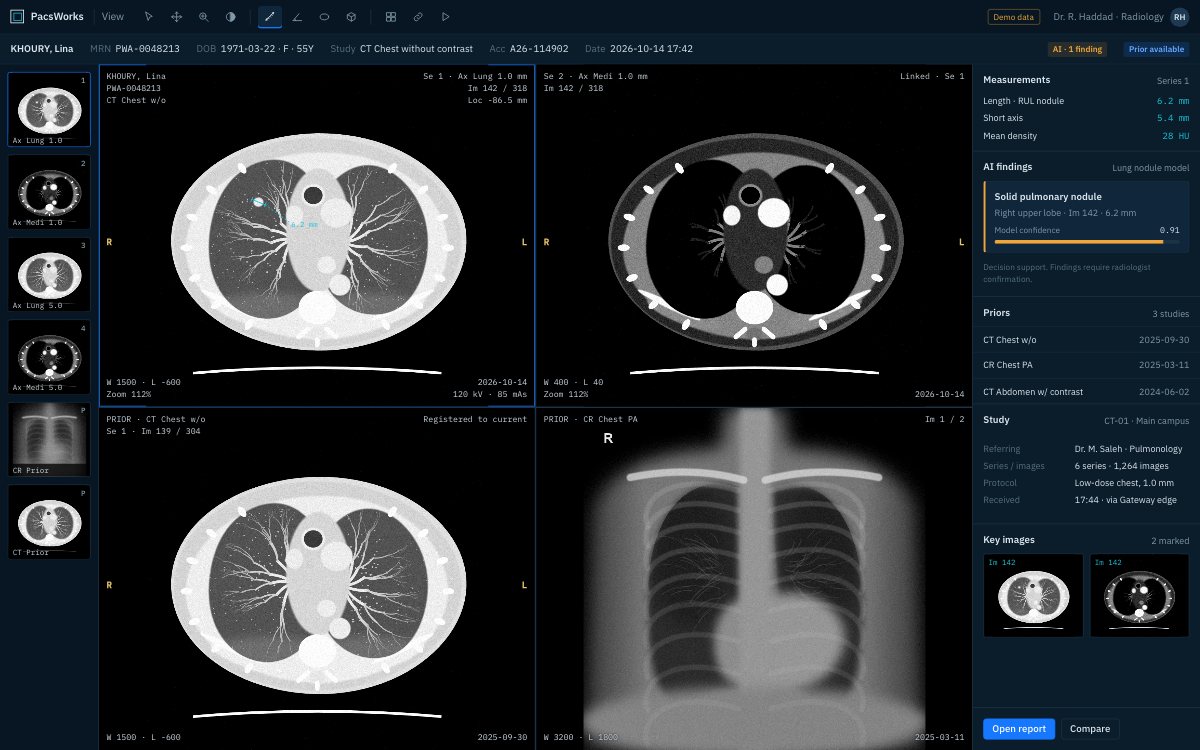
Every prior, from every department, in one viewer.
When cardiology echo, the chest CT from the satellite campus and last year’s X-ray all live in one archive, the radiologist compares them in one viewer instead of logging in to three systems.
- Current and prior studies side by side, on the same anatomy
- Hanging protocols by modality and body part
- AI overlays the radiologist confirms or rejects
Images on the ward, without a workstation.
Ward doctors, surgeons and consultants open images and the final report in a browser on a desktop, tablet or phone, launched from the patient’s record in the EMR. Nothing to install, and access follows their role.
- Launched from the EMR in context
- Desktop, tablet and phone browsers
- Every view logged for audit
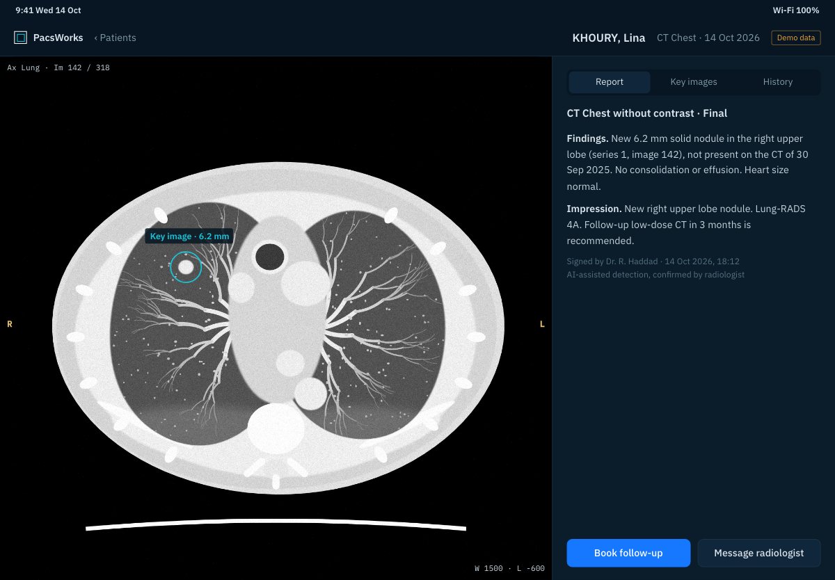
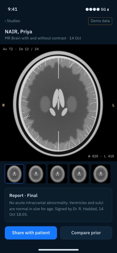
Screens show demo data and synthetic images.
What each team gets.
Radiologists
One worklist, priors ready on open, and AI results inside the viewer rather than in another window.
Referring clinicians
Images and reports from the patient record, on whatever device is at hand.
Imaging IT
One platform to patch, back up and monitor, with routing and retention defined centrally.
Hospital leadership
Fewer contracts, less duplicated storage and an archive the hospital owns independently of any viewer.
How a rollout is phased.
No single cut-over. Each phase runs alongside what is already in service.
Archive first
The vendor-neutral archive goes in beside existing PACS, which start copying studies to it.
Gateways per campus
Edge gateways take over scanner routing, so scanners stop pointing at individual systems.
Move reading
Departments move to the PacsWorks worklist one at a time, with the old system available for lookup.
Retire legacy
Old archives are migrated and reconciled, then switched off when the counts match.
Questions we hear.
Do we have to replace every PACS at once?
No. The archive and gateways go in beside what you run today, and departments move over one at a time. Each legacy system stays available until its studies have been migrated and the counts reconciled.
Can cardiology and other departments share the archive?
Yes. The archive stores standard DICOM from any department, so radiology, cardiology and other imaging services can keep one record per patient.
What happens to reading during an outage?
Reading services run on redundant servers, and a separate disaster-recovery copy of the archive is kept on a second site or in the cloud. The recovery procedure is tested as part of the service, not left on paper.
How do clinicians reach images?
From the patient’s record in the EMR. A link opens the browser viewer in context, with the images and final report the clinician’s role allows.
Can we add AI later?
Yes. AI Hub can be added at any stage. Most hospitals start with one model on one study type, such as head CT triage, and extend once it has proved itself.
Tell us about your imaging environment.
What you run today, where the bottlenecks are and how you expect to grow. We will come back with an architecture, not a product pitch.